biotech
The Physiology of HIIT
Today we look at the physiology underlying HIIT training with the aim of finding out the secret of why HIIT appears superior to many other training distributions.
info
Background
HIT stands for High-Intensity Training, or “HIIT” once you add intervals. As long as you can tolerate it, there is fairly good evidence that high-volume HIIT produces greater gains (e.g., high peak power, higher linkVO2max peak, higher FTP) than low-volume HIIT. And a training regime that includes higher amounts of HIIT is more likely to improve VO2max, neuromuscular power, anaerobic power and possible aerobic/endurance than the same time spent training at low intensity (base) or even middle intensity (threshold). So HIIT is highly effective, especially in the short term.
The question for today is how does HIIT work? Why is it more effective than threshold/sweetspot or base (low intensity) work?
There are numerous possible physiological mechanisms. We know training-induced increase in chamber size and left ventricular mass (LVM) can lead to a greater stroke volume and COmax, leading to an increase in VO2max. Increases in blood volume (BV), plasma volume, red blood cell expansion, and haemoglobin mass (HbM) are also important central factors influencing VO2max. An increase in red blood cell volume leads to an enhanced oxygen-carrying capacity of the blood. A greater plasma volume (i.e., hypervolaemia) produces an increase in venous return with subsequent increases in stroke volume, COmax, and oxygen delivery.
favorite
HIIT and Cardiovascular Benefits
Cardiovascular adaptations are not unique to HIIT at all, but may be superior to those of continuous endurance training (according to Helgerud et al., 2007; Wisløff, & Kemi, 2009) at least in short-term studies. That makes sense, HIIT is a stronger stimulus. For example, the magnitude of cardiomyocyte hypertrophy depends on the intensity of exercise and in one study HIT induced a substantially larger response than moderate intensity: specifically 14% versus 5% longer cells linkref.
Slørdahl et al. (2004) demonstrated that high-intensity aerobic training at 90–95% of maximal oxygen consumption (VO2max) increased left ventricle heart mass by 12% and cardiac contractility by 13% linkref.
Helgerud et al. link18 found a HIIT protocol conducted for eight weeks was more effective than performing the same total work in a continuous manner at lower intensities for improving VO2max, and this corresponded to an increase in stroke volume. Remember that stroke volume changes more than HRmax in response to training.
Compared to lower intensity exercise, higher intensity exercise stimulates larger increases in HR, ventilation, and body temperature and also a larger EPOC response from HIIT/SIT compared to the MICT.
fitness_center
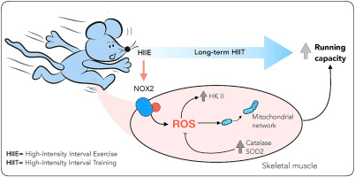
HIIT and Skeletal Muscle Adaptations
As a rule of thumb, muscle hypertrophy occurs in response to both frequency of training and training load. For example, see quadriceps muscle hypertrophy with classical motor endurance training (predicted by both the frequency of training and training load) linkKonopka and Harber 2014.
MacDougall et al. (1998) demonstrated increased skeletal muscle oxidative enzyme levels of citrate synthase (36%), malate dehydrogenase (29%), and succinate dehydrogenase (65%) among students engaging in 7 weeks of HIIT cycling sprints linkref.
Recruitment of “inefficient” fast-twitch fibres may also contribute to benefits due to the high levels of type II fibre recruitment in sprint training (SIT), subsequently leading to greater fatigue and metabolic disturbances associated with the increased ATP and/or O2 cost of exercise linkref.
bolt
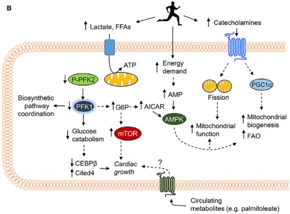
HIIT and Metabolic Adaptations
HIT is associated with greater levels of glycogen depletion as observed with maximal sprinting linkref.
Perry et al. (2008) showed that fat oxidation, or fat burning, was significantly higher and carbohydrate oxidation (burning) significantly lower after 6 weeks of interval training.
There is also the research on EPOC (Excess Post-exercise Oxygen Consumption). For a review see this linklink.
Various studies find higher EPOC with HIIT training as compared to continuous aerobic training linkref with only a couple reporting no differences. Research to date suggests small differences in EPOC post-HIIT compared to MICT in the immediate (<1 hour) recovery period, but greater EPOC values post-HIIT when examined over 24 hours and more gains with sprint intervals (SIT) than HIT or MICT.
science
HIIT and Mitochondria
The mitochondrion is the primary organelle for energy production, generating adenosine triphosphate (ATP) via the electron transport system (ETS) using substrates from the tricarboxylic acid (TCA) cycle. However, modern geroscience and exercise physiology have shifted the focus from simply how mitochondria produce energy to how specific training variables—intensity versus volume—dictate their size, efficiency, and lifecycle.
speed
The Role of Exercise Intensity
Recent research has clarified how exercise intensity drives specific functional mitochondrial adaptations. Granata et al. (2016) demonstrated this by directly comparing traditional long slow distance (LSD) training, High-Intensity Interval Training (HIIT), and Sprint Interval Training (SIT) in young, moderately trained men.
After four weeks, only the SIT group showed a 25% increase in maximal mitochondrial respiration. This functional upgrade was driven by increased protein content of PGC-1α, p53, and PHF20 (a protein that stabilises the tumour suppressor p53 to regulate mitochondrial function).
While SIT provides the most rapid changes in respiration per minute of exercise, HIIT alone remains highly effective. Earlier studies (Daussin et al., 2008; Jacobs et al., 2013) demonstrated that just two weeks of HIIT increases both mitochondrial respiration and content (measured via cytochrome c oxidase [COX] activity), leading to rapid increases in overall exercise capacity.
layers
The Role of Exercise Volume
When training intensity is maintained but total volume is manipulated, the biological response shifts from respiration efficiency to mitochondrial enlargement and biogenesis.
Granata et al. showed that increasing training volume (comparing a high-volume HIT period to a mid-volume HIT period) significantly increased both mitochondrial respiration and Citrate Synthase (CS) activity—a primary marker of mitochondrial mass/density—by approximately 50%. Notably, after two weeks of reduced training volume (detraining), respiration remained high, while CS activity began to decline, indicating that mitochondrial mass is highly sensitive to total work volume.
However, intensity still magnifies the volume effect. When total work volume is strictly matched, higher intensity yields a greater metabolic signal. MacInnis et al. (2017) found that volume-matched single-leg HIIT produced a significantly greater increase in CS activity compared to Moderate-Intensity Continuous Training (MICT) (39% vs. 11%).
autorenew
The New Frontier: Mitochondrial Quality Control (MQC)
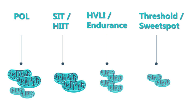
Modern physiology synthesises these findings into a unified model of mitochondrial dynamics known as Mitochondrial Quality Control (MQC). Mitochondria are not static powerhouses; they constantly remodel themselves based on the specific stress applied:
- Function over Mass (HIIT/SIT): High-intensity training provides severe metabolic and mechanical stress that triggers mitophagy—the selective cellular degradation of damaged, inefficient mitochondria. It forces mitochondrial fission (splitting), pruning defective, ROS-leaking mitochondria and replacing them with highly efficient units.
- Mass over Function (MICT/Zone 2): High-volume, moderate-intensity training maximises mitochondrial mass and network connectivity. Sustained energy demand promotes mitochondrial fusion (joining together), creating larger, highly interconnected networks optimised for long-term lipid oxidation and increased capillary density.
table_chart
Summary of Mitochondrial Adaptations
| Training Modality | Intensity Profile | Primary Cellular Target | Structural & Functional Adaptation |
|---|---|---|---|
| Moderate-Intensity Continuous Training (MICT / LSD) | 60–75% HRmax | Mitochondrial Mass / Content | Promotes mitochondrial fusion (building interconnected networks); increases Citrate Synthase (CS) activity and capillary density. |
| High-Intensity Interval Training (HIIT) | 85–95% HRmax | Mitochondrial Function & Biogenesis | Up-regulates PGC-1α and p53; increases cytochrome c oxidase (COX) activity; balances mass and respiration increases. |
| Sprint Interval Training (SIT) | >100% VO2 max | Mitochondrial Respiration & Quality Control | Intensely stimulates fission and mitophagy to clear damaged units; yields highest time-efficiency for respiratory gains. |
analytics
A Meta-analysis of Benefits
linkRosenblat and colleagues (2022) recently looked at how interval training affects physiology. They examined 32 studies (369 participants) on both HIIT (n = 18) and SIT (n = 17) interventions. There was a low sample size in inactive people and in athletes, but the result in active (non-athletes) was intriguing in terms of pre-post training change (delta).
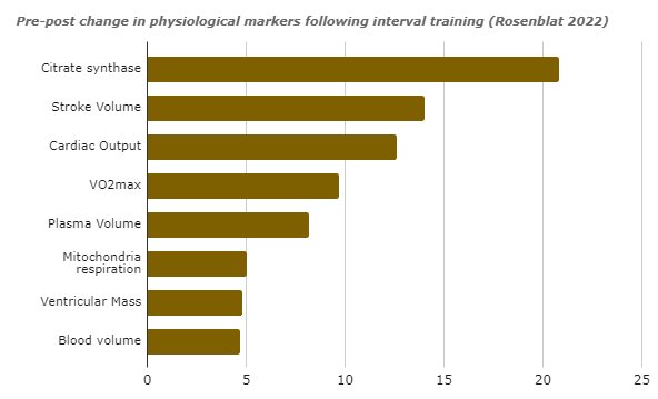
Interestingly, HIIT had larger effects than SIT. Further, there were significant effects on capillary density and VO2max in athletes.
summarize
Summary
HIT (especially HIIT) is generally more effective in the same training time (but it is significantly harder) than steady-state training.
HIIT is important for increasing mitochondrial activity, whereas a greater training volume is needed to increase mitochondrial mass.
HIT usually means a lot of intervals, but typically in a balanced plan where the TID is 50:15:35 this is not 100% intervals—that would be crazy!
There is a downside that in the long-term, high amounts of HIIT can risk overtraining and burnout, which is why POLARISED training combines HIT with base/endurance.
menu_book
Citations and Further Reading
Adaptations to Endurance and Strength Training
Excess Post-exercise Oxygen Consumption (EPOC) Review
Daussin, F. N., et al. (2008). Effect of interval versus continuous training on cardiorespiratory and mitochondrial functions. American Journal of Physiology-Regulatory, Integrative and Comparative Physiology.
Granata, C., et al. (2016). Training intensity modulates changes in PGC-1α and p53 protein content and mitochondrial respiration, but not markers of mitochondrial biogenesis in human skeletal muscle. The FASEB Journal.
Jacobs, R. A., et al. (2013). Improvements in exercise performance with high-intensity interval training coincide with an increase in skeletal muscle mitochondrial content and function. Journal of Applied Physiology.
MacInnis, M. J., et al. (2017). Superior mitochondrial adaptations in human skeletal muscle after interval compared to continuous single-leg cycling matched for total work. The Journal of Physiology.
This is part two in our HIT series. Part 1 is linkhere and Part 3 is coming soon.